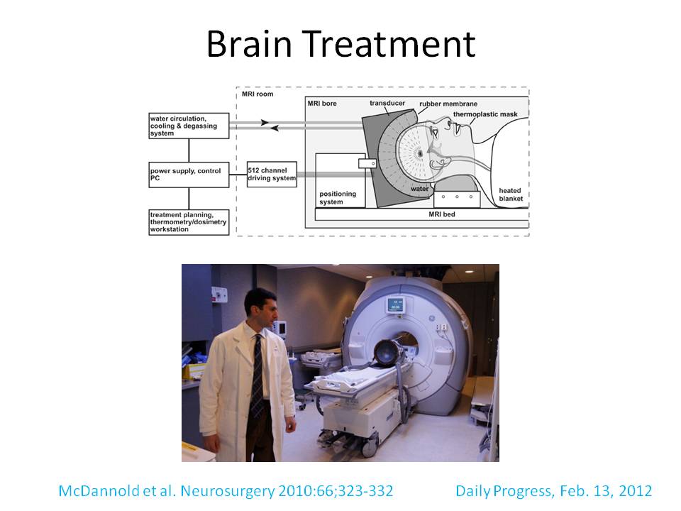
An exciting new study has launched to evaluate the use of focused ultrasound to destroy hypothalamic hamartomas. Learn more about the procedure and the study from Dr. Nathan Fountain.
Listen to an interview and see images of this new procedure below.
Hypothalamic hamartoma
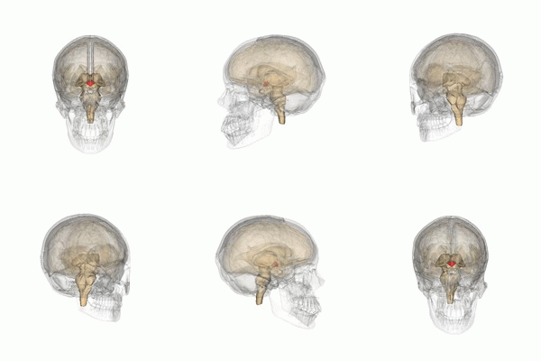
- Hypothalamic hamartoma (HH) is a rare condition where a tumor-like structure forms in the hypothalamus. The hypothalamus is a part of the brain important for production of hormones that regulate growth, blood-pressure, heart rate and hunger.
[By Images are generated by Life Science Databases(LSDB). [CC BY-SA 2.1 jp], via Wikimedia Commons]
- HHs are characterized by frequent intellectual disability and seizures that do not respond well to medication.
- Since the hamartomas are located in the hypothalamus, which is in the center of the brain, they are difficult to treat surgically.
Focused ultrasound
- An exciting new study that is currently going on aims to use focused ultrasound waves to destroy the tumor-like structure.
- The principle behind focused ultrasound is similar to using a magnifying glass to focus light to burn a hole in a piece of paper. In focused ultrasound, an (acoustic) lens is used to concentrate ultrasound waves into a spot that is deep inside the body. In this particular study, focused ultrasound will be used to destroy tissue that is located deep in the brain.
- Because of this, the technique is non-invasive (i.e. no probe physically enters the brain).
- Since this is a novel study, the main goal is to see if focused ultrasound method is safe.
The second goal is to measure its effectiveness by comparing seizure frequency before and after the procedure. - Researchers do not anticipate extreme side-effects, but one issue could be that the normal brain tissue is destroyed in addition to the malformed brain tissue. Since the hypothalamus will be targeted, this could lead to abnormal hormone production. The researchers will keep an eye on this potential issue by measuring hormone levels throughout the study.
- Focused ultrasound is being used for numerous other diseases e.g. essential tremor, prostate disease and uterine fibroids. This is the first study that will examine its usefulness in HH and similar other conditions.

Focused Ultrasound for Treatment of HH – Dr. Nathan Fountain
Who can participate? (more information here)
- Adults 18 or older
- International patients are eligible
- At least 3 seizures per month
- Currently taking 2 anti-epileptic drugs
- Previously failed 2 anti-epileptic drugs
- Evidence that shows that seizures are arising from a lesion deep inside the brain
The study
- The study is more than a year long, and procedures are at no cost to the participants. International participants are also welcome, and some transportation costs can be borne by the study.
- For questions, please contact the study coordinator, Stacy Thompson, RN SRC2H@virginia.edu or the principle investigator, Dr. Nathan Fountain NBF2P@virginia.edu.
Advantages of focused ultrasound
- The process is entirely non-invasive as ultrasound waves will be applied on the scalp.
- Patients are less likely to experience bleeding or infection (this is one way in which this technique differs from the traditional techniques to treat HH).
- Anesthesia not necessary although sedation may be used
- The area of malformation can be visualized even before the actual procedure. This will allow the researchers to be certain of the area to destroy.


